|
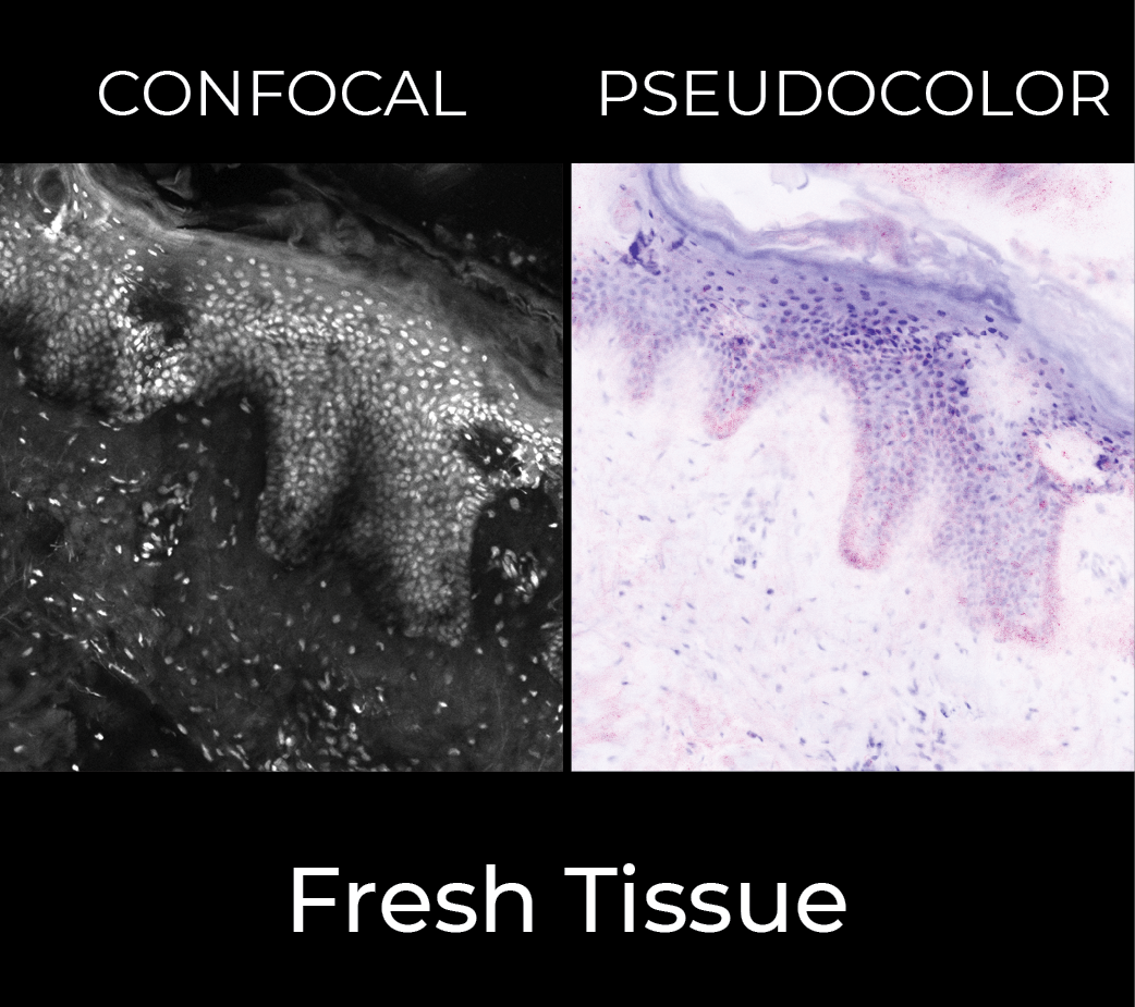
Fresh specimen imaging
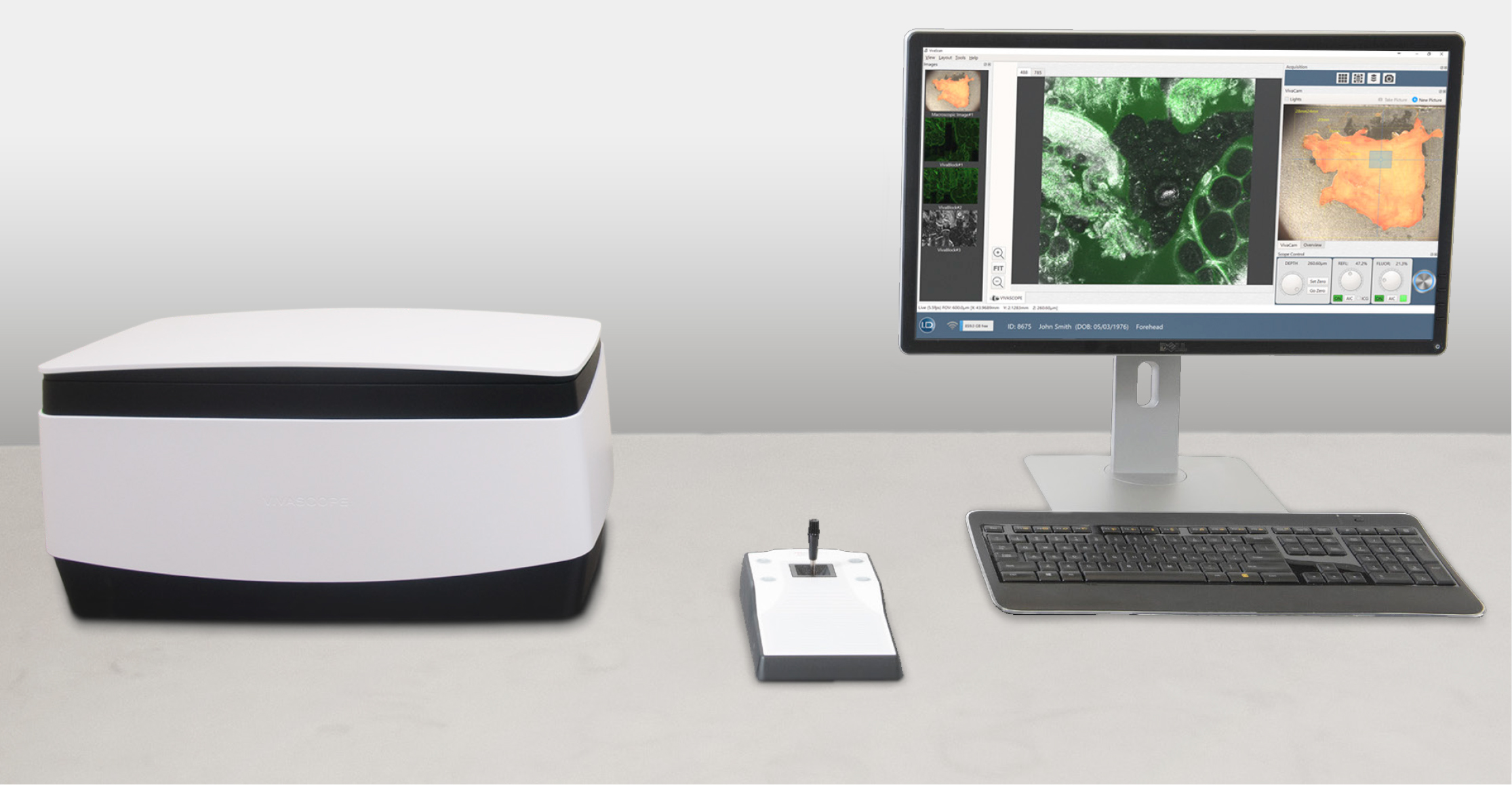
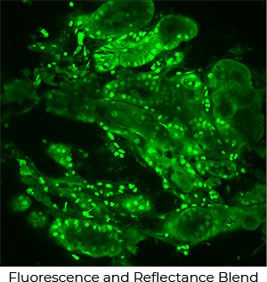
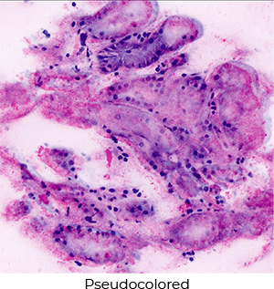
The VIVASCOPE 2500 is a confocal microscope specially designed for imaging fresh, needle aspirated, or fixed specimens in reflectance (phase contrast) mode or in fluorescence mode for specimens stained with fluorochromes. Specimens can be examined in near real-time without time consuming processing procedures. The VIVASCOPE 2500 is used by physicians and other licensed healthcare professionals to view enlarged images of specimens during pathological examinations.
Features
- Two lasers:
- 488nm (blue)
- 785 nm (infrared)
- Filter sets for fluorescent dyes including:
- Acridine orange
- Fluorescein
- Indocyanine green
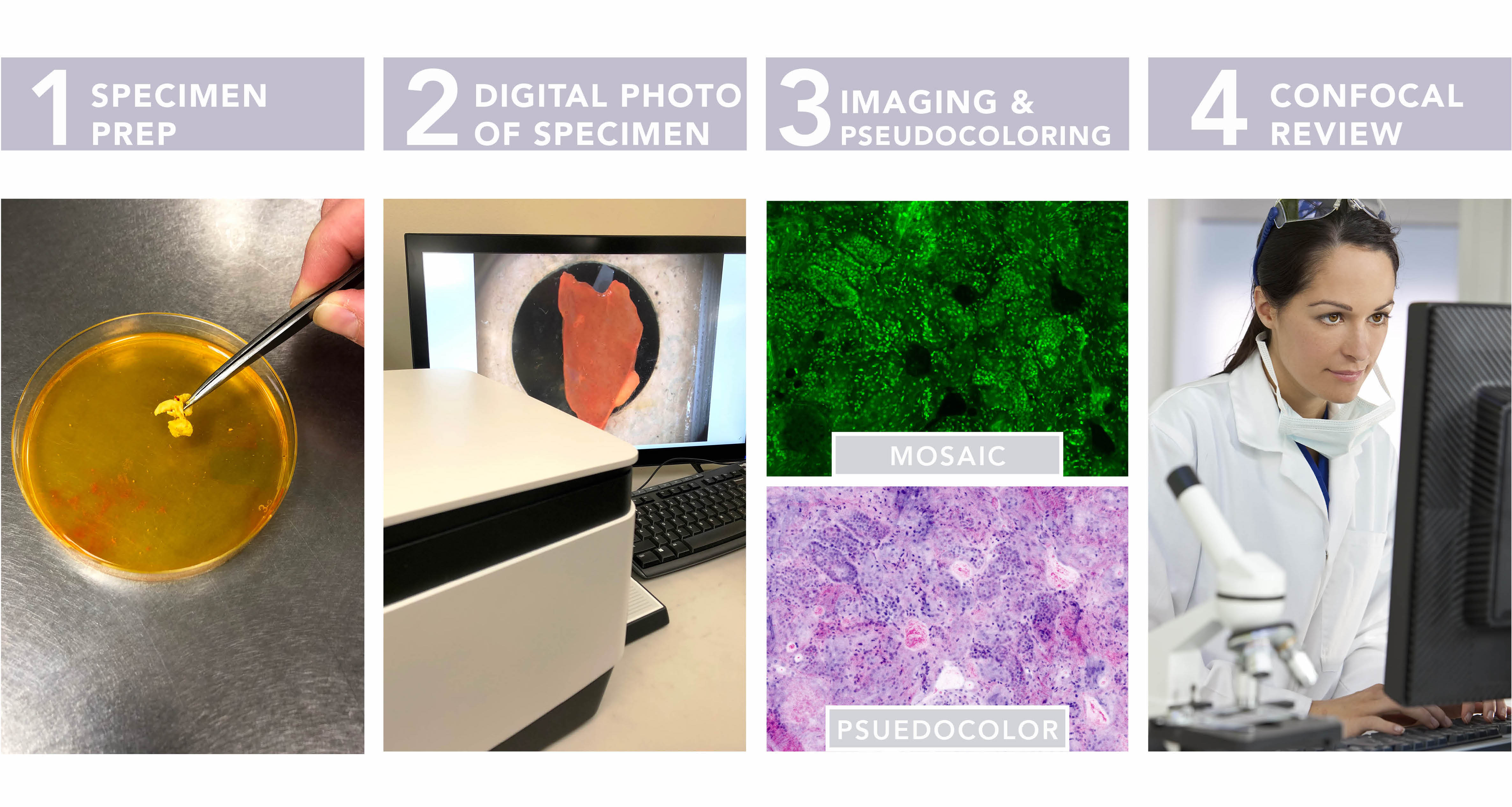
- Digital macro camera:
- Offers high-resolution photo of specimen
- Correlates to confocal image for navigation
- Simplifies selection of imaged area
Literature

| Optical Section Thickness |
<4μm |
| Single Field of View Size |
550μm x 550μm |
| Image Resolution |
1024 x 1024 pixels (single field of view) |
| Maximum Sample Size |
20 mm X 20 mm |
| Operating Wavelengths |
488 & 785nm |
| Objective |
0.9 NA Water Immersion Lens |
| Regulatory Certifications and Standards |
- FDA Class 1 Medical Device
- IEC 61010-1:2010
- IEC 61326: 2013
- IEC 60825-1: 2014
|
| Laser Classification |
|
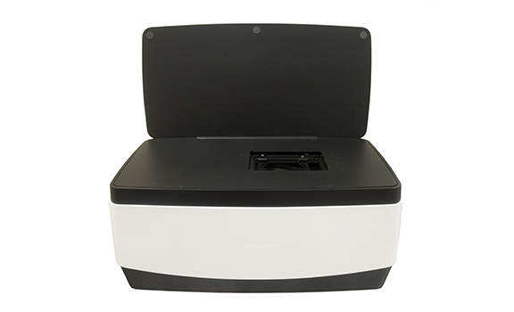
Dimensions
LxWxH |
Scan Head Only
25x52.5x25 cm |
| Weight |
17.2 kg |
| Power Source |
100-120 V~, 50-60Hz
220-240 V~, 50-60Hz |
| Operating Humidity |
Non-condensing |
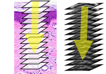
The VIVASCOPE 2500 generates single images of tissue that are parallel to the tissue surface or on the “horizontal plane” .
The image depth, or position within the specimen, is changed by moving the objective lens up, down or laterally, relative to the specimen surface. |
 |
| A series of horizontal images captured in “Z-depth”, or from the surface of the specimen to a preconfigured depth. The image plane is incrementally changed by moving the objective lens of the VIVASCOPE 2500 and images are captured at consecutively deeper depths within the tissue. |
 |
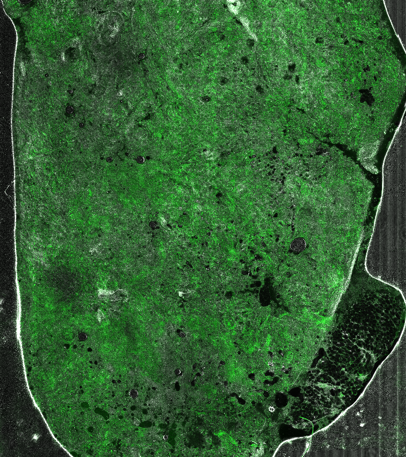
| An array of images taken on a single horizontal plane creates a mosaic. The VIVASCOPE 2500 scans the objective lens laterally over the tissue while capturing a series of images and displaying them as a composite image covering an area up to 20 x 20mm. |
 |
|
BENEFITS
- Specimen preparation in minutes
- High-resolution images in near real-time
- Image can be magnified and panned
|
|