Caliber I.D. Inc., VIVASCOPE, VIVANET, and VIVACAM are registered trademarks of Caliber I.D. Inc., or its subsidiaries and affiliated companies.
| VIVASCOPE System: Non-invasive cellular imaging of the skin |
|
|
CONFOCAL IMAGING The VIVASCOPE® system is an in vivo |
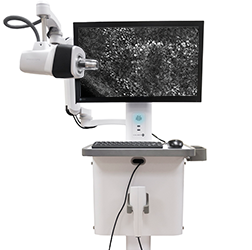
VIVASCOPE 1500: Ability to image up to an 8 x 8 mm areaThe VIVASCOPE 1500 reflectance confocal imaging system offers a non-invasive way to image the skin in vivo from the surface to the superficial collagen layers.* Highlights
Key SpecificationsMapped Field: 8 x 8 mm in both the X & Y directions Single Frame FOV: 500 µm x 500 µm Displayed Image Resolution: 1024 x 1024 pixels Depth of Imaging: Superficial collagen layers* Image Formats: Native DICOM files exportable as: BMP, PNG, JPEG, and TIFF |
 |
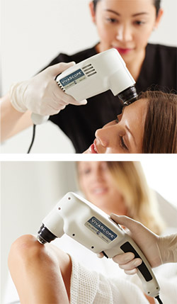
VIVASCOPE 3000: Flexible hand-held confocal imaging deviceThe VIVASCOPE 3000 is a flexible, hand-held in vivo reflectance confocal microscope for skin imaging. This imaging tool allows the operator to freely navigate the device across the skin while delivering stable, repeatable, high quality cellular-resolution images of epithelium of the skin and supporting stroma.
Highlights
Key SpecificationsSingle Frame FOV: 750 μm X 750 μm Displayed Image Resolution: 1024 X 1024 pixels Frame Rate: 6 frames per second Image Formats: Native DICOM files exportable as BMP, PNG, JPEG, and TIFF
|
 |
VIVASCANVIVASCAN software incorporates comprehensive features that allow users to acquire, transfer, display, review, and store images, in one, easy-to-use dashboard. Additionally, VIVASCAN makes it easy to schedule patients for exams, perform imaging exams on one or more areas during a single patient visit, and retrieve images associated with the patient’s history. |
 |
*depending on tissue |
Experience VIVASCOPEExamineAreas of interest are identified by
ImageCaptures high-resolution cellularimage of epithelium of the skin and the supporting stroma.
Review/CollaborateUsing the VIVANET communicationsystem, physicians can easily review or share images. |